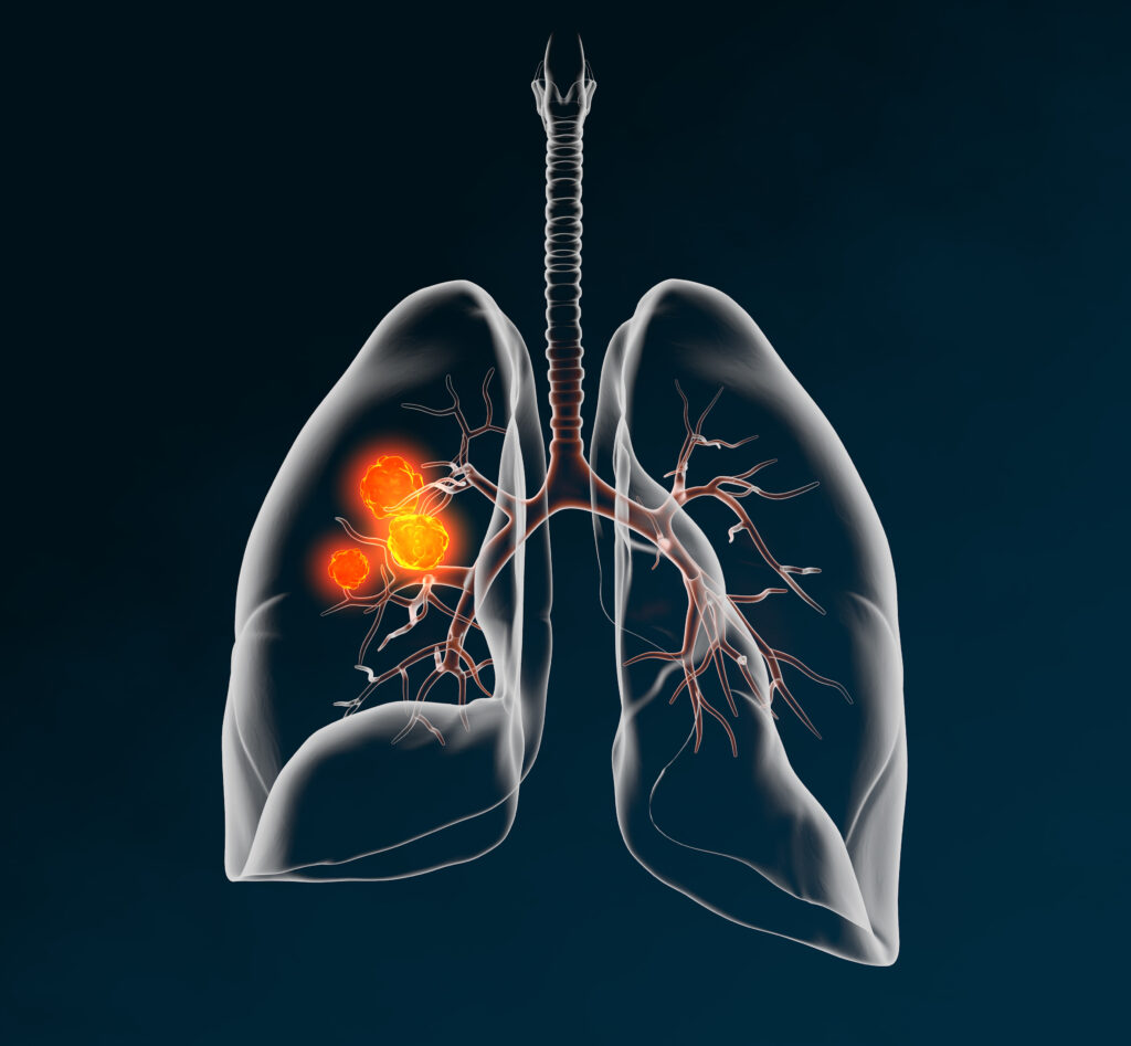
Lung cancer biopsy dislodges tumor cells into circulating blood
Aim: A “seed” of lung cancer metastasis is circulating tumor cells (CTCs), which may be dislodged from a tumor during biopsy. This possibility was assessed among patients who underwent lung tumor biopsy using flexible fiber-topic bronchoscopy (FFB).
Methods: The study involved six patients with non-small cell lung cancer who underwent FFB biopsy to diagnose a lesion pathologically (5 males and 1 female, median age 63 years, 6 adenocarcinomas, of 4 clinical-stage IA, 1 stage IB, and 1 stage IIIA), CTCs were extracted from the peripheral vein blood at pre-FFB and at post-FFB using a size selection method.
Results: No tumor cell was detected at pre- and post-FFB was in three cases (50%); no tumor cells were detected pre-FFB while CTCs were detected at post-FFB in two cases (33.3%); and CTCs were detected at pre-FFB with numerous CTCs detected at post-FFB in one case (17.7%). In addition, similar tendencies were observed in each analysis of single-cell and clustered-cell categories.
Conclusion: These results suggest that a FFB biopsy of lung cancer may potentially dislodge CTCs from a tumor into the circulating peripheral blood.
Detection of Circulating Tumour Cells and Survival of Patients with Non-small Cell Lung Cancer
Background: Detection of circulating tumour cells (CTCs) in the peripheral blood of lung cancer patients may predict survival. Various platforms exist that allow capture of these cells for further analysis; little work however, has been done with the ScreenCell device, an antibody-independent CTC platform. The aim of our study was to evaluate the ScreenCell device for detection of CTCs in lung cancer patients and to establish correlations of these findings with survival.
Materials and methods: Twenty-three patients, nine males, and fourteen females, underwent surgical treatment from February to May 2014 for non-small cell lung cancer. Thirteen patients had adenocarcinoma and ten squamous cell carcinoma, while eight were at an early stage (I-II) and five at a later stage (III-IV). Blood samples were obtained prior to surgery and following filtration through the ScreenCell device, were independently reviewed by 2 consultant pathologists.
Results: The pathologists were able to independently identify CTCs in 78.3% (N=18) and 73.9% (N=17) of the cases examined, with overall 80.6% in early stages compared to 60.0% in late stages. The median survival times of positive vs. negative for CTC patients were 1011 and 711 days respectively, with a survival percentage rate of 77.8% and 60% in positive and negative CTC cohorts respectively.
Conclusion: The results of this study suggest that the presence of CTCs analyzed by ScreenCell did not necessarily lead to a poorer prognosis in patients with lung cancer after curative surgery.
Circulating tumour cells in patients with lung cancer undergoing endobronchial cryotherapy
Early diagnosis of lung cancer still poses a major issue, with a large proportion of patients diagnosed at late stages. Therapeutic options and treatment remain limited in these patients. In most cases only palliative therapies are available to alleviate any severe symptoms. Endobronchial cryotherapy (EC) is one form of palliative treatment offered to patients with obstructive airway tumours. Although successful, the impact on circulating tumour cell (CTCs) spread has not been investigated in detail. This study recruited 20 patients awaiting EC treatment. Baseline and post EC blood samples were analysed for presence of CTCs. Results showed an increase in CTCs following EC in 75% of patients. Significant increases were noticeable in some cases. Although EC is a well-accepted modality of treatment to alleviate symptoms, it may lead to an increase in CTCs, which in turn may have implications for tumour issemination and metastatic spread.
Circulating Tumour Cells in Patients with Malignant Lung Tumors Undergoing Radio-frequency Ablation
Background/Aim: Radiofrequency ablation (RFA) is an increasingly utilised technique in patients with surgically-untreatable lesions. The effect of this therapy on circulating tumor cells (CTCs) is unknown. As far as we are aware of, this is the first study to evaluate the effects of RFA on CTCs in patients with malignant lung tumors immediately post-treatment. Patients and Methods: Nine patients with primary or metastatic lung tumors underwent RFA therapy from June to November 2013. Blood samples were taken before and after RFA, and filtered through the ScreenCell CTC capture device. Results: A general increase in CTCs in 7 out of the 9 cases was found, the largest increases were seen in the metastatic group. Conclusion: This study demonstrates that the manipulation and ablative procedure of lung tumors leads to immediate dissemination of tumor cells, the effects of which are unknown and require further investigation.
Circulating Tumor Cells in Diagnosing LungCancer: Clinical and Morphologic Analysis

Background: The purpose of this study was to evaluate the value of circulating non-hematologic cells to differentiate benign from malignant lung lesions and their comparison with clinico-histologic features of corresponding primary lesions.
Methods: Circulating cells were isolated by size method from peripheral blood of 77 patients with malignant (n = 60) and benign (n = 17) lung lesions. They were morphologically classified as cells with malignant feature; cells with uncertain malignant feature; and cells with benign feature; then statistically correlated with clinico-cytopathologic characteristics of corresponding lung lesion.
Results: Malignant circulating cells were detected in 54 of 60 (90%) malignant patients, and in 1 of 17 (5%) benign patients; benign circulating cells in 1 of 60 (1%) malignant patients and in 15 of 17 (88%) benign patients; and circulating cells with uncertain malignant aspect in 5 of 60 (8%) malignant patients and 1 of 17 (5%) benign patients. For a malignant circulating cells count greater than 25, sensitivity and specificity were 89% and 100%, respectively. The count was significantly correlated with stage, size, and standard uptake value of primary tumor. In 39 of 54 (72%) cases, the malignant circulating cells allowed a specific histologic diagnosis of the corresponding primary tumor after immunohistochemical analysis.
Conclusions: Malignant circulating cells may be a valid marker in the diagnostic workup of lung lesions. However, our resuts should be corroborated by larger future studies especially for patients having small nodules.