Long-Term Dynamics of Three Dimensional Telomere Profiles in Circulating Tumor Cells in High-Risk Prostate Cancer Patients Undergoing Androgen-Deprivation and Radiation Therapy

Patient-specific assessment, disease monitoring, and the development of an accurate early surrogate of the therapeutic efficacy of locally advanced prostate cancer still remain a clinical challenge. Contrary to prostate biopsies, circulating tumor cell (CTC) collection from blood is a less-invasive method and has potential as a real-time liquid biopsy and as a surrogate marker for treatment efficacy. In this study, we used size-based filtration to isolate CTCs from the blood of 100 prostate cancer patients with high-risk localized disease. CTCs from five time points: +0, +2, +6, +12 and +24 months were analyzed. Consenting treatment-naïve patients with cT3, Gleason 8-10, or prostate-specific antigen > 20 ng/mL and non-metastatic prostate cancer were included. For all time points, we performed 3D telomere-specific quantitative fluorescence in situ hybridization on a minimum of thirty isolated CTCs. The patients were divided into five groups based on the changes of number of telomeres vs telomere lengths over time and into three clusters based on all telomere parameters found on diagnosis. Group 2 was classified as non-respondent to treatment and the Cluster 3 presented more aggressive phenotype. Additionally, we compared our telomere results with the PSA levels for each patient at 6 months of ADT, at 6 months of completed RT, and at 36 months post-initial therapy. CTCs of patients with PSA levels above or equal to 0.1 ng/mL presented significant increases of nuclear volume, number of telomeres, and telomere aggregates. The 3D telomere analysis of CTCs identified disease heterogeneity among a clinically homogeneous group of patients, which suggests differences in therapeutic responses. Our finding suggests a new opportunity for better treatment monitoring of patients with localized high-risk prostate cancer.

Advancing Risk Assessment of Intermediate RiskProstate Cancer Patients

The individual risk to progression is unclear for intermediate risk prostate cancer patients.
To assess their risk to progression, we examined the level of genomic instability in circulating
tumor cells (CTCs) using quantitative three-dimensional (3D) telomere analysis. Data of CTCs from
65 treatment-naïve patients with biopsy-confirmed D’Amico-defined intermediate risk prostate cancer
were compared to radical prostatectomy pathology results, which provided a clinical endpoint to the
study and confirmed pre-operative pathology or demonstrated upgrading. Hierarchical centroid cluster
analysis of 3D pre-operative CTC telomere profiling placed the patients into three subgroups with different
potential risk of aggressive disease. Logistic regression modeling of the risk of progression estimated
odds ratios with 95% confidence interval (CI) and separated patients into “stable” vs. “risk of aggressive”
disease. The receiver operating characteristic (ROC) curve showed an area under the curve (AUC) of 0.77,
while prostate specific antigen (PSA) (AUC of 0.59) and Gleason 3 + 4 = 7 vs. 4 + 3 = 7 (p > 0.6) were
unable to predict progressive or stable disease. The data suggest that quantitative 3D telomere profiling
of CTCs may be a potential tool for assessing a patient’s prostate cancer pre-treatment risk.

Filtration-based enrichment of circulating tumor cells from all prostate cancer risk groups

Objective: To combine circulating tumor cell (CTC) isolation by filtration and immunohistochemistry to investigate the presence of CTCs in low, intermediate, and high-risk prostate cancer (PCa). CTCs isolated from these risk groups stained positive for both cytokeratin and androgen receptors, but negative for CD45.

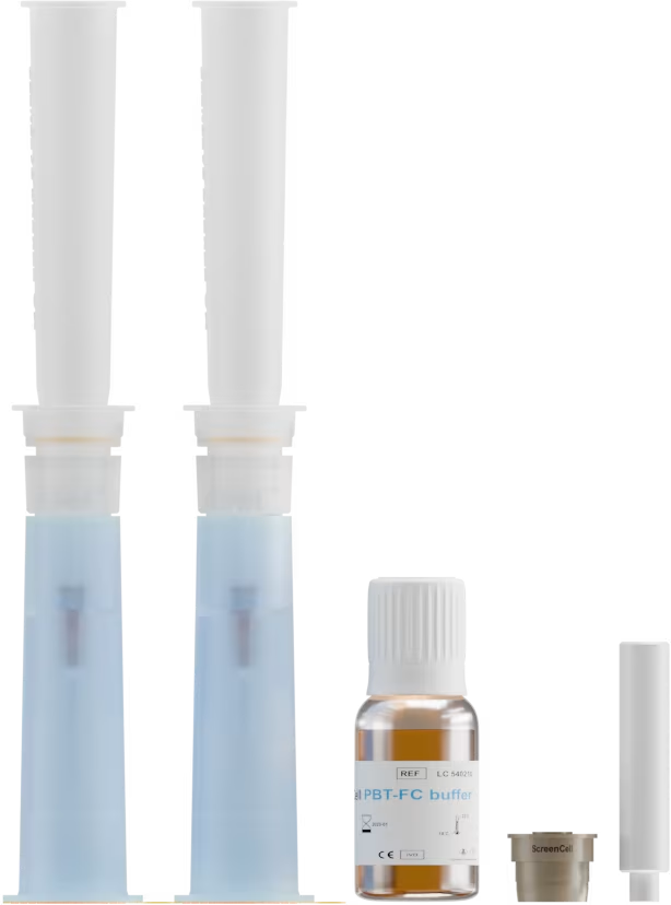
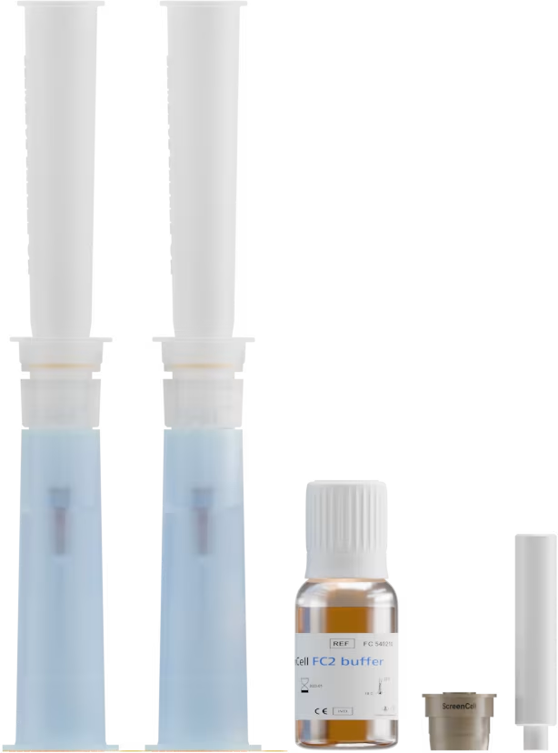
Patients and methods: Blood samples from 41 biopsy confirmed patients with PCa at different clinical stages such as low, intermediate, and high risk were analyzed. The samples were processed with the ScreenCell filtration device and PCa CTCs were captured for all patients. The isolated CTCs were confirmed PCa CTCs by the presence of androgen receptors and cytokeratins 8, 18, and 19 that occurred in the absence of CD45 positivity. PCa CTC nuclear sizes were measured using the TeloView program.

Results: The filtration-based isolation method used permitted the measurement of the average nuclear size of the captured CTCs. CTCs were identified by immunohistochemistry in low, intermediate, and high-risk groups of patients with PCa.

Conclusion: CTCs may be found in all stages of PCa. These CTCs can be used to determine the level of genomic instability at any stage of PCa; this will, in the future, enable personalized patient management.

Single-cell analysis of circulating tumor cells identifies cumulative expression patterns of EMT-related genes in metastatic prostate cancer

Background: Prostate tumors shed circulating tumor cells (CTCs) into the blood stream. Increased evidence shows that CTCs are often present in metastatic prostate cancer and can be alternative sources for disease profiling and prognostication. Here we postulate that CTCs expressing genes related to epithelial-mesenchymal transition (EMT) are strong predictors of metastatic prostate cancer.

Methods: A microfiltration system was used to trap CTCs from peripheral blood based on size selection of large epithelial-like cells without CD45 leukocyte marker. These cells individually retrieved with a micromanipulator device were assessed for cell membrane physical properties using atomic force microscopy. Additionally, 38 CTCs from eight prostate cancer patients were used to determine expression profiles of 84 EMT-related and reference genes using a microfluidics-based PCR system.

Results: Increased cell elasticity and membrane smoothness were found in CTCs compared to noncancerous cells, highlighting their potential invasiveness and mobility in the peripheral circulation. Despite heterogeneous expression patterns of individual CTCs, genes that promote mesenchymal transitioning into a more malignant state, including IGF1, IGF2, EGFR, FOXP3, and TGFB3, were commonly observed in these cells. An additional subset of EMT-related genes (e.g., PTPRN2, ALDH1, ESR2, and WNT5A) were expressed in CTCs of castration-resistant cancer, but less frequently in castration-sensitive cancer.

Conclusions: The study suggests that an incremental expression of EMT-related genes in CTCs is associated with metastatic castration-resistant cancer. Although CTCs represent a group of highly heterogeneous cells, their unique EMT-related gene signatures provide a new opportunity for personalized treatments with targeted inhibitors in advanced prostate cancer patients.

Three-Dimensional Telomeric Analysis of Isolated Circulating Tumor Cells (CTCs) Defines CTC Subpopulations

Circulating tumor cells (CTCs) have been identified with the potential to serve as suitable biomarkers for tumor stage and progression, but the availability of effective isolation technique(s) coupled with detailed molecular characterization have been the challenges encountered in making CTCs clinically relevant. For the first time, we combined isolation of CTCs using the ScreenCell filtration technique with quantitative analysis of CTC telomeres by TeloView. This resulted in the identification and molecular characterization of different subpopulations of CTCs in the same patient. Three-dimensional (3D) telomeric analysis was carried out on isolated CTCs of 19 patients that consisted of four different tumor types, namely, prostate, colon, breast, melanoma, and one lung cancer cell line. With telomeric analysis of the filter-isolated CTCs, the level of chromosomal instability (CIN) of the CTCs can be determined. Our study shows that subpopulations of CTCs can be identified on the basis of their 3D telomeric properties.