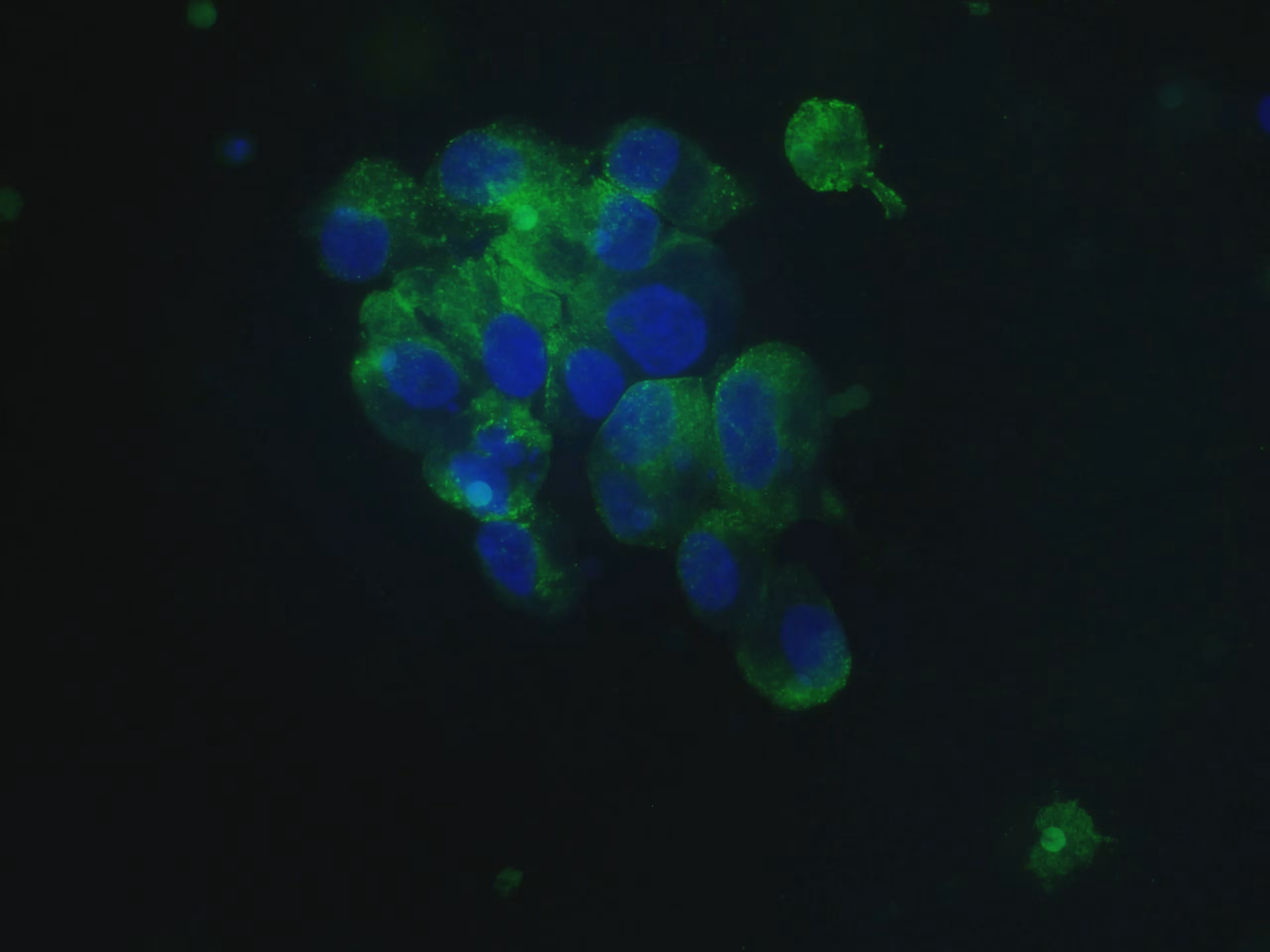

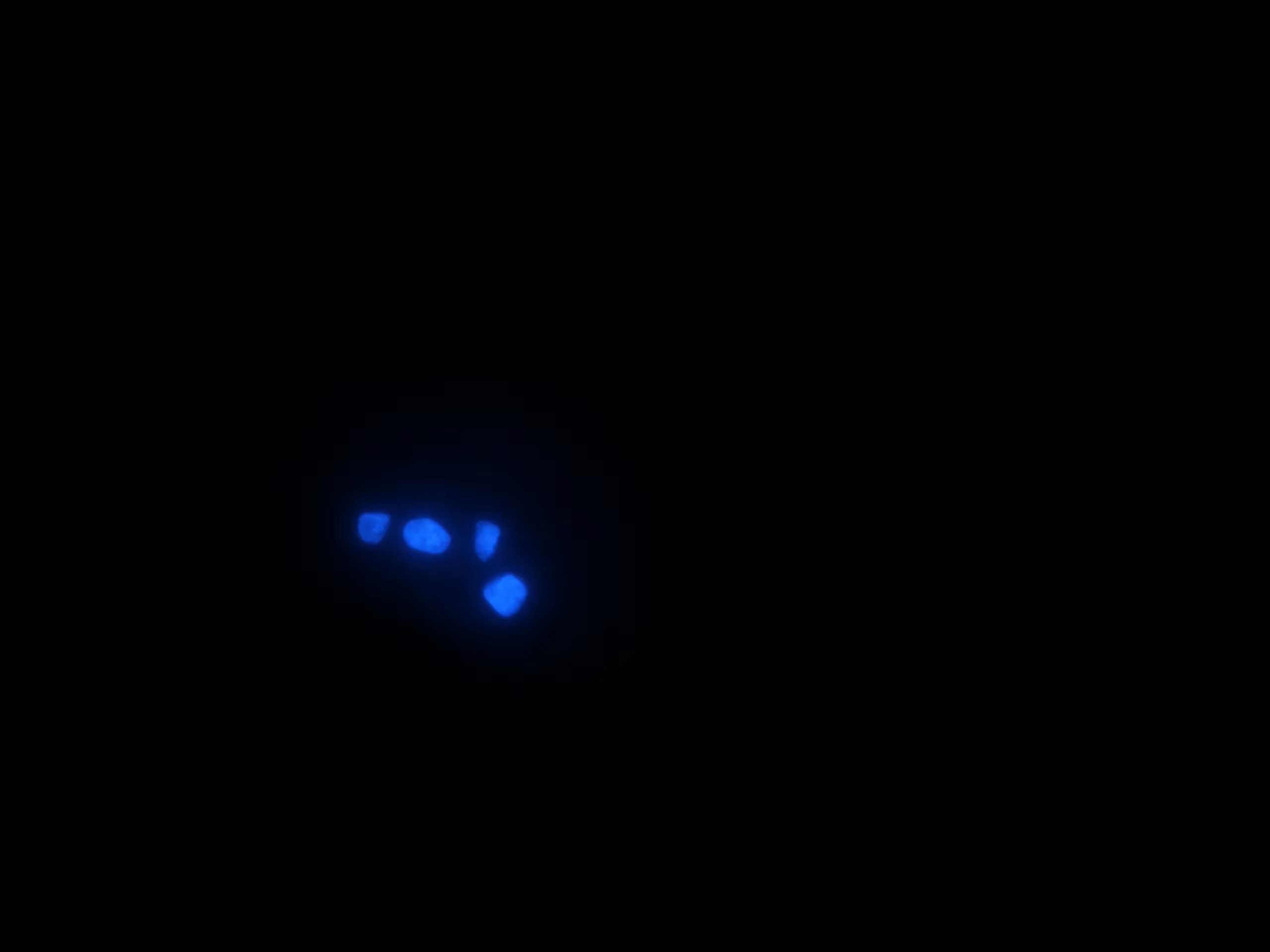
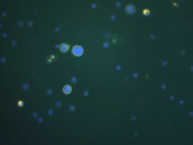
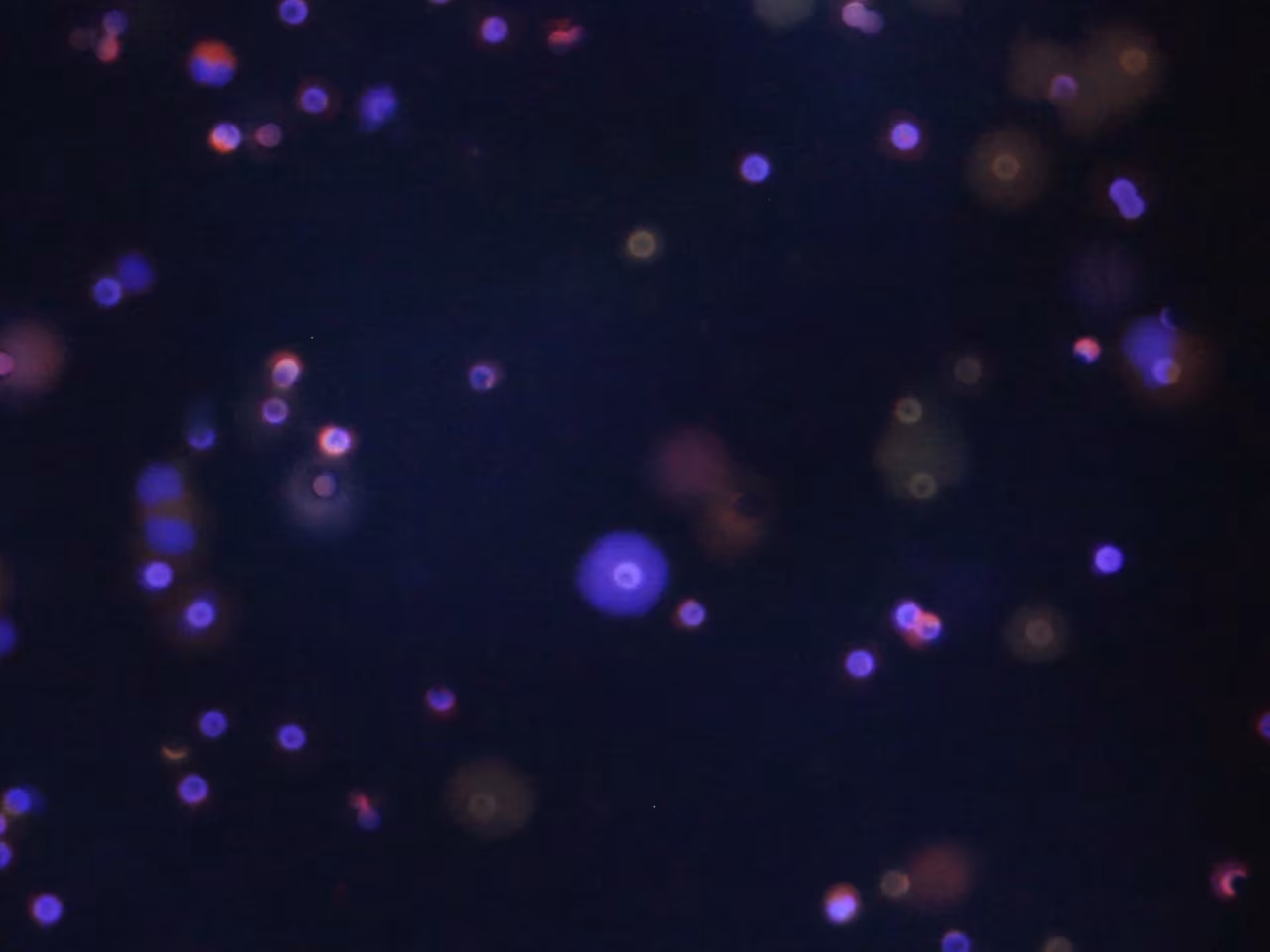
Immunofluorescence staining on cancer cells is an essential technique in oncology because it allows the multiplexed analysis of several cellular markers simultaneously. This ability to detect several specific biomarkers in a single preparation offers a detailed and comprehensive view of CTCs, thus facilitating the study of their heterogeneity and the evaluation of their role in tumor evolution.
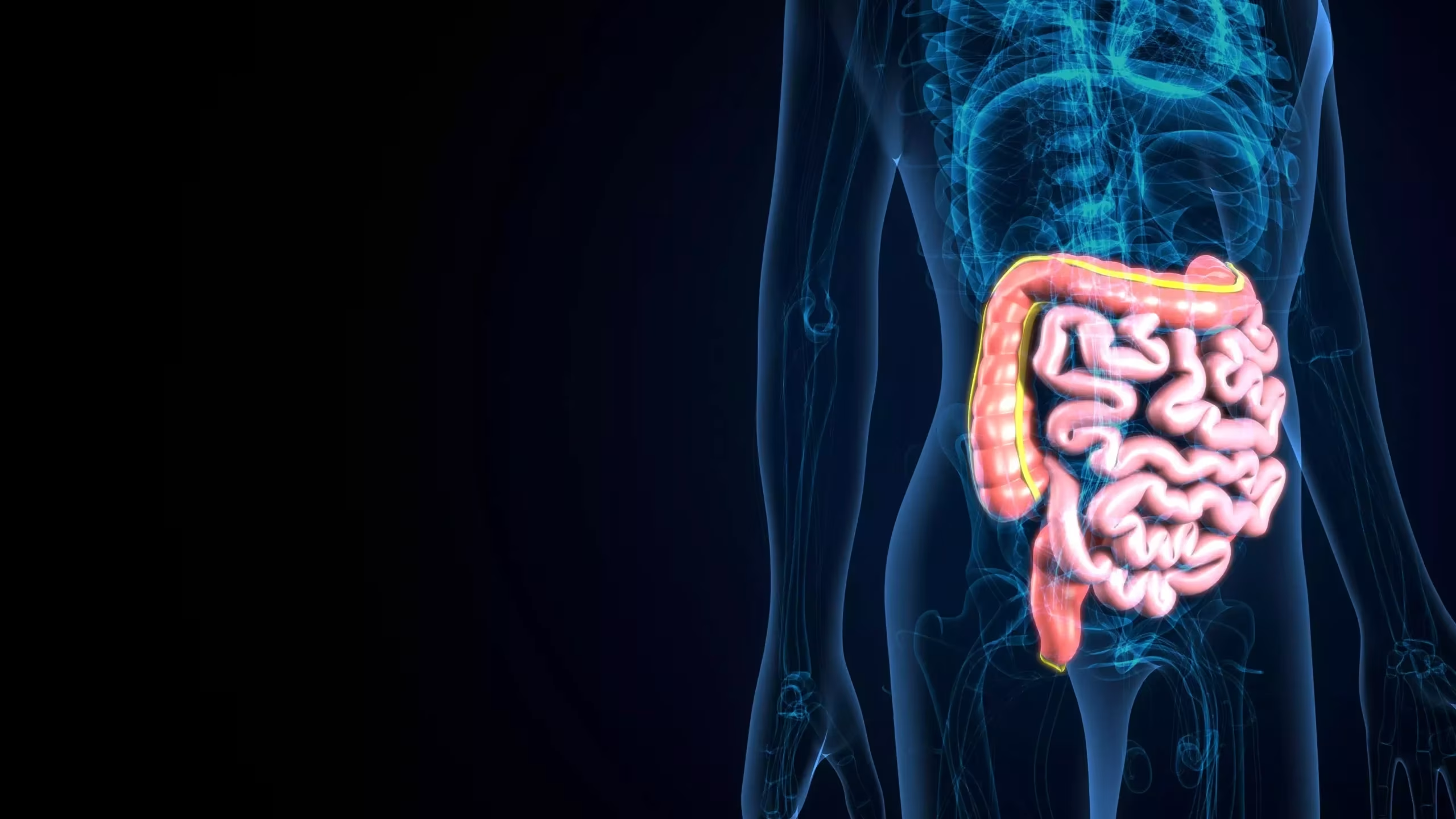
HT29 colorectal cancer cell line
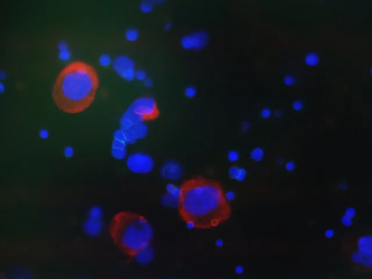
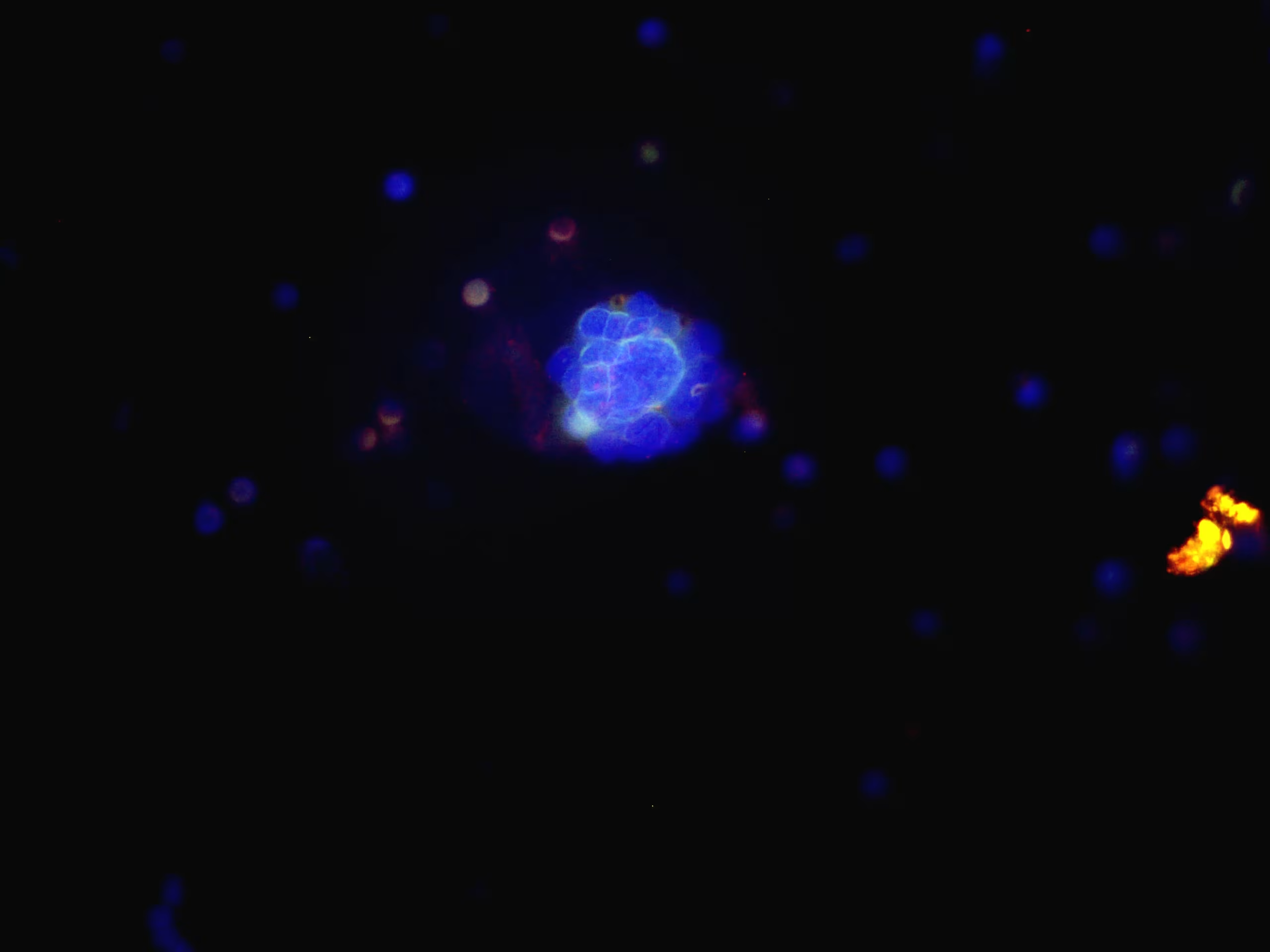
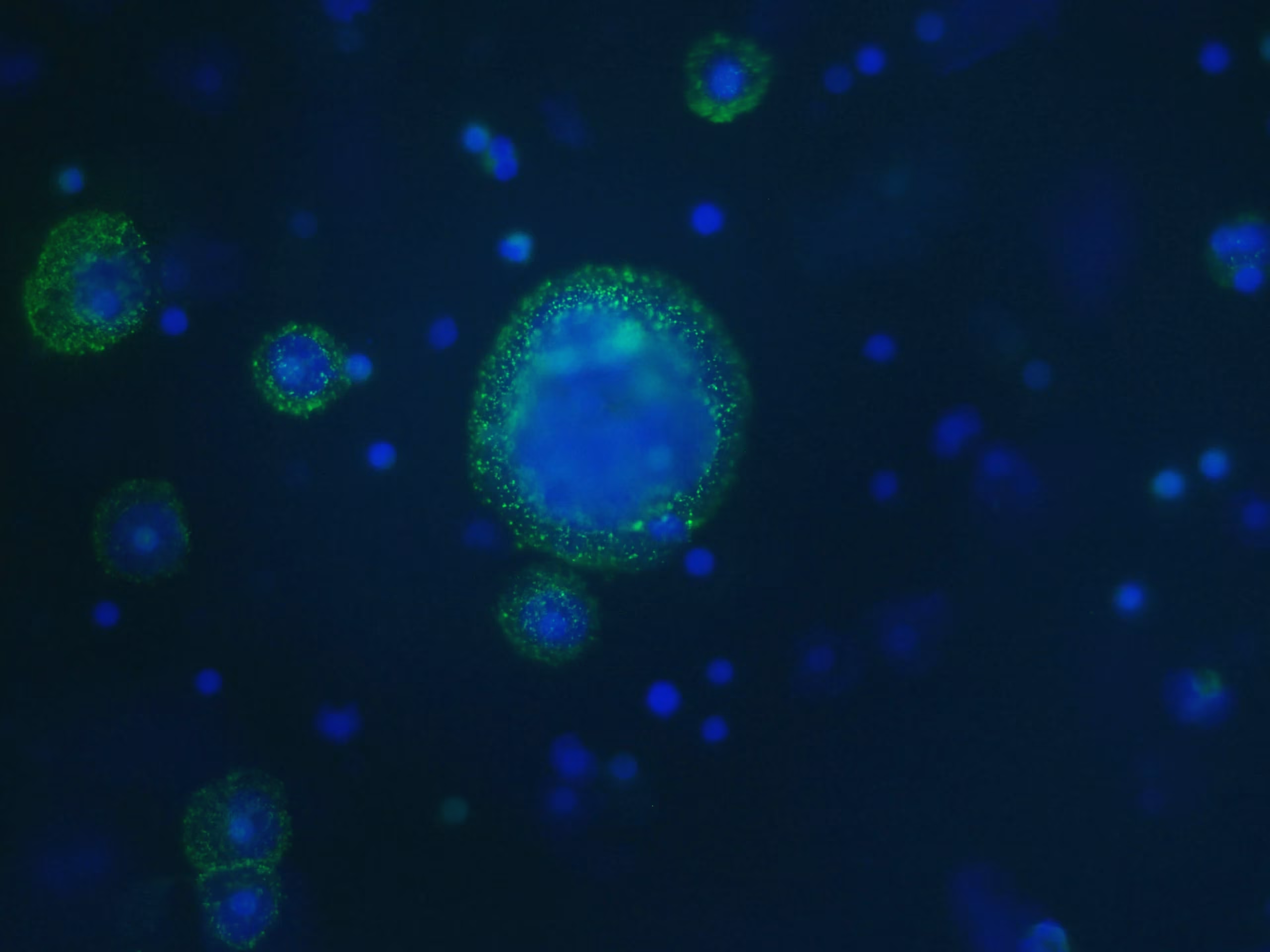
Immunofluorescence staining on CTCs is a crucial tool to explore the molecular characteristics of circulating tumor cells. It provides detailed information on the surface profiles of CTCs, facilitating the identification of specific tumor cells in the blood.
“Results from immunofluorescence experiments demonstrated that, overall, EpICD and/or EpEX was expressed in 176 CTCs detected by ScreenCell, while the CellSearch® system was able to capture only 10 CTCs.”
Ref : EpCAM-expressing circulating tumor cells in colorectal cancer – DOI: 10.5301/ijbm.5000284
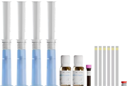
Blood sample
< 4 hours
Cells fixed and analyzed on the isolation support
Cytological
staining
Immunofluorescence
Immunocytochemistry
Fluorescence in situ hybridization
Blood sample
< 3 days
Cytological
staining
Immunofluorescence
Immunocytochemistry
Fluorescence in situ hybridization
Blood sample
< 3 days
ou
Cells fixed detached
from the isolation support for analysis in suspension or on slide
Cytological
staining
Immunofluorescence
Immunocytochemistry
Fluorescence in situ hybridization
Blood sample
< 4 hours
ou
Cells fixed detached
from the isolation support for analysis in suspension or on slide
Cytological
staining
Immunofluorescence
Immunocytochemistry
Fluorescence in situ hybridization
Blood sample
< 4 hours
Living cells detached
from the isolation support for suspension analysis
Cytological
staining
Immunofluorescence
Immunocytochemistry
Fluorescence in situ hybridization

Blood sample < 3 days
or
Fixed cells detached
from the isolation support for analysis in suspension or on slide
Blood sample < 4 hours
or
Fixed cells detached
from the isolation support for analysis in suspension or on slide
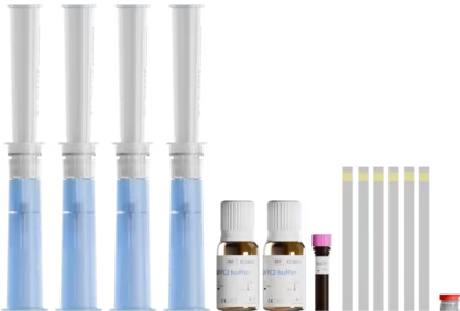
Yes, immunofluorescence staining can be performed directly on the CYTO isolation media after CTC capture. CYTO ScreenCell® filters are specially designed to enable downstream staining procedures without cell transfer, thus preserving morphology and avoiding loss.
We strongly recommend using our protocols, specifically developed and optimized by our team. These protocols are designed to ensure reliable, high-quality results in your CTC immunofluorescence staining experiments.
To simplify your workflow, we offer optimized, ready-to-use protocols validated for immunofluorescence applications on our isolation media.
Are you interested in establishing a fundamental or clinical research project focused on circulating tumor cells (CTCs) or liquid biopsy ?
Share your project details with us, and we will be delighted to assist you in bringing it to fruition.